Equipment
Adapt Dx dark adaptation
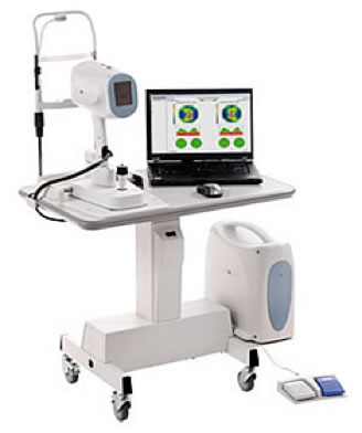
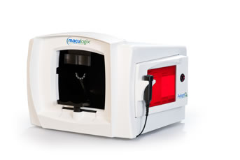 Dark adaptation testing is used to monitor age-related macular degeneration and any other condition that may cause night blindness. Imagine you awake in the middle of the night to go to the bathroom. You turn on the light in the bathroom, and then when you are done and need to return to bed, you turn out the light. In people that have macular degeneration, that sudden change from a bright environment to a dark environment takes longer for the eyes to adapt to the darkness. Testing can find out how long it takes for the eyes to adapt. During the testing, a bright light is flashed, and then a button is pushed when you see a second light appear.
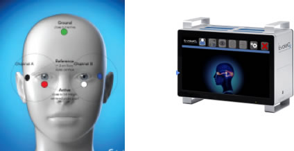
Evoke Dx VEP testing
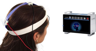
Evoke Dx ERG testing
Rebound Tonometry (RBT)
Color Dx
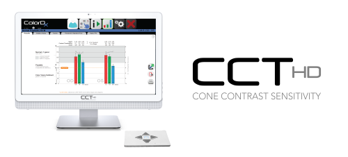 ColorDx is an advanced way of checking color vision. It will tell the doctor the specific type of deficiency a “color blind” person may be facing. Color vision testing is also used to monitor the progression of certain diseases or to watch for unwanted side effects of certain medications. ColorDx involves the use of a tablet to perform color vision testing.
InflammaDry
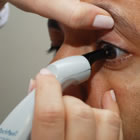
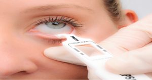 InflammaDry is a diagnostic test that can be performed in your eye doctor’s office to test your tears for the presence of inflammation. Inflammation may increase as dry eye progresses into a chronic condition, and if left untreated, can potentially damage the cells on the surface of your eye. By using the InflammaDry test to determine if there is an above normal level of inflammation on the surface of your eye, your eye doctor can determine the best and most appropriate dry eye treatment plan for you.
A small, soft piece of fabric will be gently dabbed along your lower eyelid to collect tears, very similar to the way a paper towel absorbs liquid from a surface. The tear collection process takes less than a minute and is not painful. Once the tear sample is collected, the InflammaDry test is activated and results are provided before you leave your doctor’s office. A positive InflammaDry test result indicates that there is a significant amount of inflammation on the surface of the eye.
Tear Lab
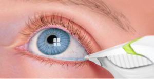 Tear Lab is a device that measures the osmolarity of tears. If tears evaporate too quickly, they can leave behind a salt residue which makes the eyes more uncomfortable. During testing, a small device is used to collect a sample of your tears to measure the osmolarity.
Fishbaugh Family Eyecare is the only eyecare provider in Mercer and Allen counties to offer this technology

Corneal topographer
 Corneal topography is an easy way for the doctor to get a detailed, visual description of the surface of the cornea. A machine will scan the front of your eye, and nothing will touch your eye. The results will help the doctor to determine if you have irregular astigmatism or other conditions such as keratoconus. Topography can also be helpful in certain contact lens fittings.
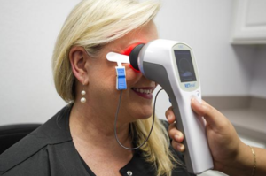
Optical Coherence Tomography
 Optical Coherence Tomography (OCT) is a machine that scans the eye painlessly. During testing, you will be asked to look at a target, and the machine will do a scan. Nothing will touch your eye. OCT testing can be used to help monitor glaucoma, macular degeneration, narrow anterior chamber angles, or any other condition that may affect the optic nerve or macula.
Fundus Photography/Anterior Segment Photography
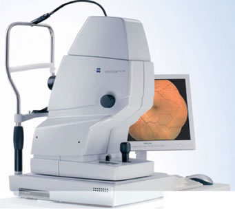 Our offices are equipped with special cameras that allow photographs to be taken on either the inside or the outside of the eyes. Photographs are useful to help the doctor monitor conditions for changes over time.
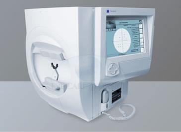
Automated Visual Field Machines
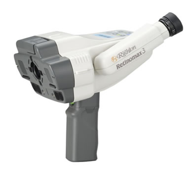
Auto Refractor
Pachymetry
Full-field Electroretinogram
Full-field electroretinogram (ffERG) allows doctors to monitor changes in the eyes due to diabetic retinopathy, glaucoma, or other retinal diseases. During testing an electrode is placed on the skin near the eye. A hand-held device is then placed in front of the eye. A screen will flicker for 3-5 minutes to get results for each eye.
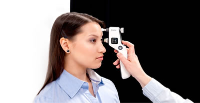
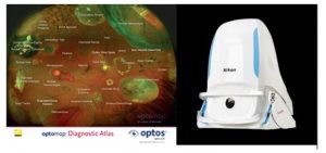
Optomap
The optomap ultra-wide digital retinal imaging device captures more than 80% of your retina in one image. The retina is the only place in the body where blood vessels can be seen directly. With this image, the doctor can detect even the earliest sign of disease that appears on the retina. Early detection is essential so treatments can be administered.
Come See the Difference at Fishbaugh Family Eyecare
Contact us today, we are ready to answer your questions. Schedule your appointment at one of our two locations.
